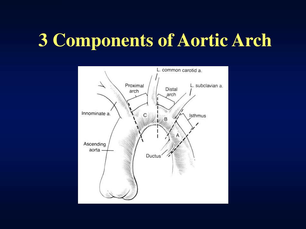

EtiologyĪlmost 50% of patients with interrupted aortic arch (IAA) have a 22q11.2 deletion this cause of 22q11.2 deletion syndrome, also known as DiGeorge syndrome. Besides a ventricular septal defect, IAA can be associated with other more complicated cardiac anomalies for example, transposition of the great arteries, truncus arteriosus, aortopulmonary window, single ventricle, aortic valve atresia, right-sided ductus, and double-outlet right ventricle. Due to this malalignment, there could be left ventricular outflow tract obstruction. This lesion is present is approximately 73% of all cases. There is posterior malalignment of the conal septum additional to the interrupted aortic arch, producing a ventricular septal defect as an associated lesion. IAA is a ductus dependent lesion since this is the only way the blood flow can travel to places distal to the disruption. In an IAA, there is an anatomical and luminal disruption between the ascending and descending aorta. Interrupted aortic arch is an anomaly that can be considered the most severe form of aortic coarctation. Approximately 97% of babies born with a non-critical congenital heart disease have a life expectancy of one year of age, and approximately 95% are expected to live around 18 years of age.Ī rare type of congenital heart disease is an interrupted aortic arch (IAA), which affects approximately 1.5% of congenital heart disease patients. The survival rate of patients with congenital heart disease will depend on the severity, time of diagnosis, and treatment. the congenital heart disease represents approximately 4.5% of all neonatal deaths.

In the United States, congenital heart disease affects 1% of births (40,000) per year, of which 25% have critical Congenital heart disease. It has an incidence of 8 cases of every 1000 live birth worldwide. Explain modalities to improve care coordination among interprofessional team members in order to improve outcomes for patients affected by interrupted aortic arch.Ĭongenital heart disease is an abnormal formation of the heart or blood vessels next to the heart.Summarize the treatment of interrupted aortic arch.Review the evaluation of an infant with.This activity reviews the presentation of the interrupted aortic arch and highlights the role of the interprofessional team in its management. Interrupted aortic arch is an anomaly that can be considered the most severe form of aortic coarctation. As the pathological studies confirmed, the aortic arch was interrupted behind the left common carotid artery (type B of the interruption).A rare type of congenital heart disease is an interrupted aortic arch (IAA), which affects approximately 1.5% of congenital heart disease patients. The image 2 shows color Doppler sagittal scan of the aortic arch, clearly depicting the interruption of the aorta. Images 1, 2: The image 1 represents a sagittal scan of the fetal aorta, showing its interrupted part (arrow). Pathological studies confirmed the diagnosis of the interrupted aortic arch and interventricular septal defect with the aorto-pulmonary shunt (Image 6), and also discovered a hypoplastic thymus (Image 7). The neonate (male, 2500 g, Apgar 6, 7) was delivered at 34 weeks, but has died soon after delivery due to the cardiac failure. Our ultrasound examination revealed several anomalies of the fetus including the interrupted aortic arch, type B, (Images 1, 2), ventricular septal defect (Image 3), and aorto-pulmonary shunt (images 4,5). A 30-year-old woman first time presented to our department at 30 weeks of pregnancy.


 0 kommentar(er)
